Overview & Anatomy
Mallet finger and jersey finger represent the two ends of the distal extensor and flexor tendon injury spectrum at the distal interphalangeal (DIP) joint. Both result from forced passive movement against active muscle contraction, but in opposite directions. Mallet finger is the most common closed tendon injury in sport, and jersey finger — though less common — carries a significant risk of missed diagnosis and permanent disability if not treated promptly.
- The terminal extensor tendon inserts into the dorsal base of the distal phalanx; the flexor digitorum profundus (FDP) inserts into the volar base of the distal phalanx — both are vulnerable to avulsion-type injuries at their insertions
- The ring finger (fourth digit) is most commonly involved in jersey finger; the middle or ring finger is most commonly involved in mallet finger; the little finger is the most common digit for mallet in some series
Mallet Finger
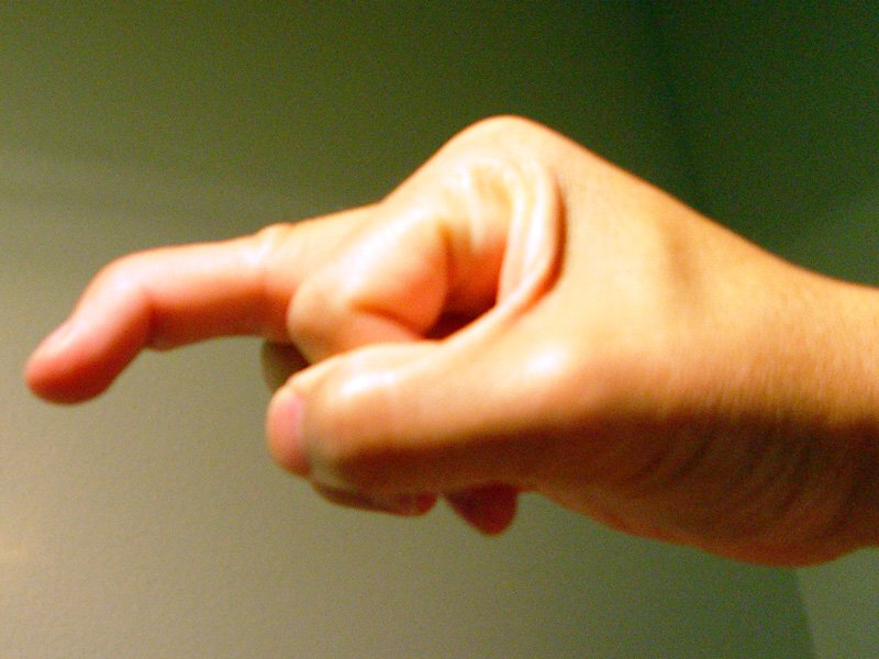
Mallet finger results from disruption of the terminal extensor tendon at or near its insertion into the distal phalanx, causing an inability to actively extend the DIP joint. It is almost always a closed injury.
- Mechanism: forced DIP flexion against an actively extending finger — classically a ball striking the fingertip (cricket, basketball, volleyball); the terminal tendon ruptures or avulses its bony insertion
- Doyle classification: Type I — closed tendon rupture with or without small avulsion fragment (most common — treat non-operatively); Type II — open laceration at or proximal to the DIP joint; Type III — deep abrasion with loss of skin, subcutaneous tissue, and tendon substance; Type IV — mallet fracture (further subdivided): IVA transepiphyseal fracture (children), IVB fracture with 20–50% of articular surface, IVC fracture with >50% articular surface or volar subluxation of distal phalanx
- Clinical features: flexed DIP joint ("drooped finger"); inability to actively extend DIP; DIP rests in flexion; tender over the dorsal DIP joint; passive extension is possible; PIP joint is not affected (distinguishes from boutonnière deformity)
| Type | Injury | Management |
|---|---|---|
| Type I (most common) | Closed tendon rupture ± small fragment | Stack splint (or aluminium foam splint) with DIP in full extension for 6 weeks continuous, then 2 weeks night-only; DIP must never flex during splinting period — even one episode of flexion restarts the 6-week clock |
| Type II | Open laceration | Wound repair + splinting; primary tendon repair if substance is adequate; K-wire across DIP in extension post-repair |
| Type III | Skin and tendon loss | Wound cover (local flap or skin graft) + DIP pin in extension; formal tendon reconstruction delayed |
| Type IVB | Bony mallet (20–50% articular surface) | Usually non-operative with splinting; surgery for significant displacement or articular step-off >2 mm |
| Type IVC | Bony mallet >50% articular surface ± volar subluxation of distal phalanx | Operative fixation if joint subluxation present — volar subluxation is the key indication; extension block K-wire pinning (Ishiguro technique); open ORIF with mini-screw or pull-out wire |
- Non-operative treatment of mallet finger: DIP extension splinting for 6 weeks continuous + 2 weeks night-only; patient compliance is paramount — the most common reason for failure is non-compliance; the splint should hold the DIP in full extension (or slight hyperextension) but not flex the PIP; the patient should be instructed to never allow the DIP to flex during the 6-week continuous period — any accidental flexion restarts the clock
- Mallet finger swan neck deformity: if untreated, a mallet finger can progress to a swan neck deformity over months to years — the terminal tendon loss allows the extensor mechanism to retract proximally, increasing tension on the central slip and causing PIP hyperextension; this is why prompt recognition and treatment is important
- Ishiguro technique (extension block K-wiring): K-wire placed through the dorsal skin proximal to the fragment (blocks the fragment from displacing dorsally during reduction), then a second K-wire pins the DIP joint in extension; allows fragment reduction and joint stabilisation without open surgery; preferred for most Type IVC mallet fractures
Jersey Finger
Jersey finger is avulsion of the flexor digitorum profundus (FDP) tendon from its insertion on the volar base of the distal phalanx. The name reflects the classic mechanism — a player grabs an opponent`s jersey as the finger is forcibly extended. It is a less common but potentially more serious injury than mallet finger because of the risk of proximal tendon retraction, vascular compromise, and permanent loss of DIP flexion if treatment is delayed.
- Mechanism: forced DIP extension against actively flexing FDP — the FDP is avulsed from its distal phalangeal insertion; the ring finger is most commonly affected (in approximately 75% of cases) — the ring finger`s FDP tendon is the weakest and the digit is typically the longest at full extension
- Clinical features: inability to actively flex the DIP joint (the defining feature); the DIP rests in extension; pain and tenderness on the volar aspect of the DIP and finger; often a missed diagnosis — the patient may present late complaining of persistent pain and weakness
- Testing for jersey finger: ask the patient to flex the DIP joint while the PIP is held in extension (to isolate FDP function from FDS); inability to flex the DIP = FDP avulsion confirmed; compare with the contralateral digit
| Leddy & Packer Type | Tendon Retraction | Blood Supply | Urgency | Management |
|---|---|---|---|---|
| Type I | Retracts into the palm — both vincula disrupted | Both vincula disrupted — no blood supply; tendon ischaemic and contracts rapidly | URGENT — repair within 7–10 days; tendon becomes non-reparable after this | Surgical repair: retrieve tendon from palm; route through A2 pulley; repair to distal phalanx via pull-out suture or bone anchor |
| Type II | Retracts to the level of the PIP joint — long vinculum intact | Long vinculum intact — adequate blood supply maintained; tendon remains viable | Less urgent — repair within 4–6 weeks | Surgical repair: tendon retrieved from PIP level; repair to distal phalanx |
| Type III | Large bony fragment avulsed; remains at A4 pulley level — does not retract further | Blood supply maintained; bony fragment prevents retraction past A4 | Less urgent — repair within 4–6 weeks | ORIF of bony fragment to distal phalanx; repair within weeks acceptable |
| Type IV | Bony avulsion + simultaneous avulsion of tendon from bony fragment (two-level injury) | Variable | Urgent | Fix bony fragment + repair tendon to fragment; technically complex |
- Type I urgency: the tendon has retracted into the palm with both vincula disrupted — there is no blood supply to the tendon; without blood supply, the tendon contracts and becomes fibrotic, making repair technically impossible; surgery must be performed within 7–10 days of injury; delayed presentation beyond 3 weeks with Type I injury has a poor prognosis for primary repair — two-stage tendon reconstruction (silicone rod + tendon graft) or DIP arthrodesis are the alternatives
- Vincula: paired blood vessels supplying the flexor tendons within the sheath — vinculum longum and vinculum breve for both FDS and FDP; disruption of both vincula in Type I jersey finger causes tendon ischaemia and rapid tendon contraction
Consultant-Level Considerations
- Missed jersey finger: one of the most common missed diagnoses in hand trauma; the patient often presents with "finger sprain" and the diagnosis is only made weeks later when they report inability to grip; by this time, Type I injuries are often irreparable; all patients with "finger sprain" should have DIP flexion tested as part of the examination; clinical suspicion in the right mechanism should prompt X-ray (to look for bony avulsion) and hand surgery review
- Late presentation jersey finger options: Type I >3–4 weeks — primary repair usually not possible due to tendon contraction and fibrosis; options: (1) DIP arthrodesis — the most reliable option for function if the patient does not need DIP flexion (provides stable painless grip); (2) Two-stage tendon reconstruction (Hunter rod + staged graft) — only if the patient has a strong functional need for DIP flexion and good pulley system
- Mallet fracture decision-making: the main surgical indication for bony mallet (Type IVC) is volar subluxation of the distal phalanx — not simply fragment size; a large fragment without subluxation can often be managed non-operatively with splinting; the Ishiguro technique is the most widely used minimally invasive method and avoids the complications of open fixation (skin necrosis over the DIP, infection, nail plate injury)
- Splint complications in mallet finger: pressure sores and skin necrosis under the Stack splint are recognised complications — the dorsal skin over the DIP is thin and at risk; check regularly; if blistering or skin breakdown develops, a brief period off the splint for skin care may be necessary but increases the risk of non-union; custom thermoplastic splints reduce this risk
Exam Pearls
- Mallet finger: forced DIP flexion → terminal extensor rupture; drooped DIP; non-operative = Stack splint × 6 weeks continuous + 2 weeks night; any accidental flexion = restart clock
- Doyle Type I (closed rupture): most common; splint; Type IVC (bony mallet >50% + volar subluxation): Ishiguro K-wire or ORIF; subluxation is the surgical indication — not fragment size alone
- Jersey finger: forced DIP extension → FDP avulsion; ring finger most common (75%); inability to flex DIP with PIP held in extension
- Leddy-Packer classification: Type I = palm retraction; both vincula disrupted; URGENT (repair within 7–10 days or irreparable)
- Type II: retracts to PIP; long vinculum intact; viable; repair within 4–6 weeks; Type III: bony fragment at A4 pulley; ORIF
- Vincula: blood supply to flexor tendons; Type I disrupts both → ischaemia and contraction → non-reparable if delayed
- Late Type I jersey finger (>3–4 weeks): DIP arthrodesis or two-stage tendon reconstruction; primary repair not possible
- Missed jersey finger: most common missed hand diagnosis; test DIP flexion in all "finger sprains"; Type I irreparable after 3 weeks
- Mallet → swan neck deformity: untreated mallet causes extensor retraction → PIP hyperextension over months; prompt treatment prevents this
- Ishiguro technique: K-wire blocks dorsal fragment; second K-wire pins DIP in extension; minimally invasive for Type IVC bony mallet