
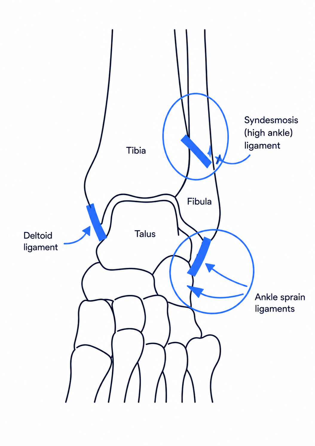
Overview — Syndesmotic Injuries
The distal tibiofibular syndesmosis is a ligamentous complex that stabilizes the relationship between the distal tibia and fibula and maintains congruence of the ankle mortise during loading. Syndesmotic injury may occur as an isolated ligament injury ("high ankle sprain") or in association with ankle fractures, particularly pronation-external rotation and pronation-abduction patterns, Weber C fractures, Maisonneuve injuries, and selected Weber B fractures.
The clinical problem is not simply whether the syndesmotic ligaments are injured, but whether the distal tibiofibular relationship is unstable. Stable injuries can usually be treated non-operatively, whereas unstable syndesmotic disruption requires accurate reduction and stabilization to restore ankle mechanics and reduce the risk of chronic pain, instability, stiffness, and post-traumatic arthritis.
- High-yield definition: a syndesmotic injury is disruption of one or more structures of the distal tibiofibular ligament complex, with treatment determined primarily by whether the syndesmosis is stable or unstable.
- Associated fractures do not automatically prove syndesmotic instability; the syndesmosis should be assessed after restoration of fibular length, rotation, and posterior malleolar anatomy.
- Anatomic reduction is more important than the choice of fixation implant.
- Malreduction of the fibula within the tibial incisura may lead to altered contact mechanics and poor outcome even when fixation is mechanically strong.
Anatomy of the Distal Tibiofibular Syndesmosis
The syndesmosis is a dynamic ligamentous articulation rather than a rigid joint. During ankle dorsiflexion, the wider anterior portion of the talus enters the mortise and produces small physiological motion of the distal fibula. The ligament complex resists excessive fibular translation, external rotation, and diastasis.
| Structure | Location / Function | Clinical Relevance |
|---|---|---|
| AITFL | Anterior inferior tibiofibular ligament; resists external rotation and anterior fibular displacement | Commonly the first ligament injured in external-rotation trauma |
| Interosseous ligament | Distal thickening of the interosseous membrane; provides central restraint | Injury may extend proximally in high-energy or Maisonneuve patterns |
| PITFL | Posterior inferior tibiofibular ligament; strong posterior restraint | May remain attached to a posterior malleolar fragment |
| Inferior transverse ligament | Deep posterior component forming a labrum-like restraint | Contributes to posterior stability of the mortise |
| Interosseous membrane | Connects tibia and fibula proximally | Proximal extension of injury is important in Maisonneuve fractures |
- Exam pearl: the posterior malleolus and PITFL are part of the syndesmotic stability complex; posterior malleolar fixation can contribute to restoration of syndesmotic stability.
Mechanism of Injury
The classic mechanism is external rotation of the talus within the ankle mortise, frequently combined with dorsiflexion. The talus acts as a wedge, forcing the distal fibula laterally and externally rotating it relative to the tibia. The injury may progress from the AITFL to the interosseous ligament and then to posterior or medial stabilizers depending on severity.
- External rotation with the foot planted is the most characteristic mechanism.
- Dorsiflexion increases stress because the wider anterior talar dome enters the ankle mortise.
- Pronation-external rotation injuries may produce deltoid injury, syndesmotic disruption, and a high fibular fracture.
- Always palpate the entire fibula when syndesmotic injury is suspected to avoid missing a Maisonneuve fracture.
- A proximal fibular fracture plus medial ankle injury should be assumed to represent syndesmotic disruption until proven otherwise.
Classification — Stable vs Unstable
Multiple grading systems exist, but for treatment decisions the most clinically useful distinction is whether the syndesmosis remains mechanically stable. The ESSKA-AFAS consensus recommends classifying acute isolated syndesmotic injuries as stable or unstable.
| Pattern | Typical Findings | General Management |
|---|---|---|
| Stable injury | Ligament injury without pathological diastasis or instability under appropriate stress/loading | Non-operative treatment |
| Latent / subtle instability | Normal or near-normal static radiographs but abnormal stress examination, weight-bearing imaging, arthroscopy, or intra-operative testing | Treat according to demonstrated instability and associated injury |
| Frankly unstable injury | Syndesmotic widening, talar shift, unstable stress test, or instability after fracture fixation | Reduction and operative stabilization |
Clinical Assessment
Clinical examination should assess the site of tenderness, mechanism, ability to bear weight, pain with external rotation, and signs of associated deltoid or proximal fibular injury. No single bedside test is sufficiently reliable to exclude syndesmotic injury; findings should be interpreted together with imaging.
| Test | Technique | Positive Finding | Pearl |
|---|---|---|---|
| AITFL tenderness | Palpate directly over the distal anterior tibiofibular joint | Focal pain over syndesmosis | Useful screening finding in acute injury |
| Squeeze test | Compress tibia and fibula together proximally at mid-calf | Pain reproduced distally at syndesmosis | Specific when positive, but a negative test does not exclude injury |
| External rotation stress test | Stabilize leg and externally rotate foot relative to tibia | Pain at distal syndesmosis / medial ankle | Reproduces common injury mechanism |
| Fibular translation test | Translate distal fibula anteriorly and posteriorly relative to tibia | Excess translation or pain compared with opposite side | Recommended in consensus assessment |
| Cotton test | Translate talus / distal fibula laterally within mortise | Excessive lateral translation or pain | Also useful intra-operatively after fracture fixation |
- Assess medial tenderness and deltoid integrity.
- Palpate proximally along the interosseous membrane and proximal fibula.
- Neurovascular examination is essential, especially with proximal fibular injuries.
- Clinical tests diagnose suspicion; instability determines treatment.
Plain Radiographic Assessment
Initial imaging should include AP, mortise, and lateral ankle radiographs, with full-length tibia-fibula imaging when a Maisonneuve injury is suspected. Radiographic measurements can support the diagnosis but are affected by positioning, rotation, and individual anatomy. Borderline values should therefore not be interpreted in isolation.
| Measurement | Where Measured | Common Reference Value | Interpretation |
|---|---|---|---|
| Tibiofibular clear space (TFCS) | Approximately 1 cm above tibial plafond on AP / mortise | < 6 mm | Widening suggests syndesmotic disruption |
| Tibiofibular overlap (TFO) | Approximately 1 cm above plafond | > 6 mm AP and > 1 mm mortise are commonly cited | Reduced or absent overlap may suggest diastasis but has anatomic variability |
| Medial clear space (MCS) | Between medial malleolus and medial talus on mortise view | Usually ≤ 4 mm | Widening suggests talar shift / deltoid incompetence and possible unstable mortise |
- Compare with the contralateral ankle when anatomy is borderline or rotational positioning is uncertain.
- Stress radiographs may uncover instability not visible on routine non-weight-bearing films.
- Normal static radiographs do not reliably exclude a clinically important syndesmotic injury.
- Do not operate on a number alone: assess the entire mortise, fibular length and rotation, medial clear space, injury pattern, and dynamic stability.
CT, MRI, Weight-Bearing CT and Arthroscopy
| Modality | Best Use | Limitation / Pearl |
|---|---|---|
| CT | Assess fibular position in incisura, posterior malleolus, occult fracture, postoperative reduction | Axial relationship is more sensitive than plain radiography; contralateral comparison may help |
| MRI | Direct visualization of AITFL, PITFL, interosseous ligament and associated cartilage / tendon injury | Excellent for ligament injury, but structural disruption does not automatically equal mechanical instability |
| Weight-bearing CT | Subtle dynamic malalignment under physiological load | Increasing role where available |
| Arthroscopy | Direct assessment of syndesmotic disruption and associated intra-articular pathology | Useful when diagnosis or instability remains uncertain |
Intra-operative Assessment of Syndesmotic Stability
In ankle fractures, syndesmotic stability should generally be tested after the major osseous components have been anatomically reduced and fixed. Failure to restore fibular length or rotation can create an apparent syndesmotic abnormality that cannot be corrected reliably by simply tightening a syndesmotic implant.
- Restore fibular length, alignment, and rotation first.
- Address the posterior malleolus when indicated; fixation may restore tension through the PITFL.
- Assess the mortise and medial clear space fluoroscopically.
- Use the Cotton / hook test or external rotation stress test to assess residual instability.
- Compare with the contralateral side if uncertainty persists.
- Key surgical principle: syndesmotic fixation should maintain an anatomic reduction; it should not be used to pull a malreduced fibula into an assumed position.
Indications for Operative Fixation
Operative stabilization is indicated when the distal tibiofibular syndesmosis is mechanically unstable or remains unstable after fixation of associated fractures.
- Frank syndesmotic diastasis on imaging.
- Persistent widening or instability on intra-operative stress testing after anatomic fracture fixation.
- Unstable isolated syndesmotic injury demonstrated by stress imaging, weight-bearing evaluation, arthroscopy, or other validated assessment.
- Maisonneuve injury with unstable distal syndesmosis.
- Selected fracture-dislocation patterns with residual syndesmotic instability after restoration of fibular and posterior malleolar anatomy.
- Stable isolated syndesmotic injuries are generally treated non-operatively; unstable injuries require operative stabilization.
Reduction of the Syndesmosis
The most important step in syndesmotic surgery is accurate reduction of the fibula within the tibial incisura. Both over-compression and rotational malreduction are possible. Reduction should be confirmed using multiple fluoroscopic views and, where available or indicated, direct visualization or CT-based assessment.
- Correct fibular shortening and malrotation before syndesmotic reduction.
- A reduction clamp may be applied across the distal tibia and fibula, but clamp position influences the direction of reduction.
- Excessive clamp force can over-compress the syndesmosis.
- Direct visualization through an anterolateral approach may improve confidence in difficult cases.
- Postoperative CT can identify malreduction not apparent on plain radiographs.
- The implant does not compensate for poor reduction. Accurate fibular position in the incisura is the goal.
Syndesmotic Screw Fixation
Trans-syndesmotic screw fixation is a traditional and effective method of stabilizing an unstable distal tibiofibular syndesmosis. The screw rigidly links the fibula to the tibia while the injured ligament complex heals.
| Technical Variable | Common Practice / Evidence | Practical Point |
|---|---|---|
| Screw diameter | 3.5 mm or 4.5 mm cortical screws are commonly used | Clinical superiority of one diameter is not clearly established |
| Number of screws | One screw is common; two screws provide greater biomechanical stability | Two screws are often considered in proximal fibular / Maisonneuve patterns, high instability, or poor bone quality |
| Cortices | Tricortical or quadricortical fixation | Clinical studies have not consistently shown a meaningful outcome difference |
| Level | Commonly approximately 2–4 cm proximal to tibial plafond | Avoid excessively distal placement through the joint region |
| Direction | Placed from fibula toward tibia, generally parallel to the ankle joint in the coronal plane and directed anteromedially according to local anatomy | Aim perpendicular to the intended tibia-fibula relationship rather than relying on a fixed universal angle |
- Advantages: inexpensive, widely available, familiar technique, strong rigid fixation.
- Limitations: restricts physiological syndesmotic motion, screw loosening or breakage, possible need for secondary removal, and risk of malreduction if fixation is performed in a non-anatomic position.
- Exam pearl: one vs two screws, 3 vs 4 cortices, and 3.5 vs 4.5 mm are technical choices; none is as important as anatomic reduction.
Suture-Button / Dynamic Fixation
Suture-button devices use cortical buttons connected by high-strength suture or tape to stabilize the syndesmosis while permitting a degree of physiological micromotion. They are often referred to as dynamic fixation devices.
- Potential advantages include preservation of physiological motion and lower routine implant-removal rates.
- Dynamic fixation may allow earlier progression of rehabilitation in selected protocols.
- Potential complications include soft-tissue irritation, infection around the lateral button, osteolysis, tunnel enlargement, button subsidence, and improper button seating.
- Correct reduction remains essential; a suture-button can also hold the syndesmosis in a malreduced position.
- One or more devices may be used depending on instability pattern, bone quality, associated fracture pattern, and surgeon preference.
- Suture-button fixation is dynamic, not "self-reducing": the syndesmosis must still be accurately reduced before final tensioning.
Screw vs Suture-Button Fixation
| Feature | Syndesmotic Screw | Suture-Button |
|---|---|---|
| Fixation concept | Rigid trans-syndesmotic fixation | Dynamic cortical suspension |
| Physiological motion | More restricted while intact | Allows controlled micromotion |
| Implant cost | Lower | Higher |
| Need for removal | Removal may be needed for symptoms, but routine removal is increasingly avoided | Usually retained unless symptomatic |
| Hardware failure | Loosening / breakage may occur | Button irritation, tunnel problems, suture failure are possible |
| Malreduction | Possible | Possible |
| Overall evidence | Reliable traditional option | Many contemporary studies show comparable or better functional / reoperation profiles, but implant choice remains case-dependent |
- Several systematic reviews and trials report similar or improved functional outcomes and fewer implant-removal procedures with suture-button constructs.
- Differences between studies include fracture patterns, device generations, screw-removal protocols, rehabilitation, and methods used to assess reduction.
- Practical conclusion: both methods can work well. Choose the fixation that permits reliable anatomic reduction, adequate stability, and a rehabilitation plan appropriate for the patient and fracture pattern.
When Should a Syndesmotic Screw Be Removed?
Historically, syndesmotic screws were routinely removed after healing because of concern that an intact screw would restrict ankle motion, break during weight bearing, or cause pain. Contemporary evidence has challenged the need for routine removal.
| Strategy | Rationale | Current Interpretation |
|---|---|---|
| Routine removal | Traditionally around 8–12 weeks or later after presumed ligament healing | Not routinely necessary in asymptomatic patients based on contemporary randomized evidence |
| On-demand removal | Remove only for clinically meaningful symptoms or another specific indication | Supported by randomized evidence and avoids an unnecessary second procedure in many patients |
| Early removal before healing | May theoretically improve motion but risks recurrent diastasis if ligament healing is incomplete | Should not be performed simply because a fixed postoperative date has been reached |
- Potential indications for removal include persistent local hardware pain, prominent implant, infection, restricted motion clearly attributable to the screw, or another planned procedure.
- Screw breakage alone is not automatically an indication for removal if the patient is asymptomatic and reduction is maintained.
- Removal itself carries risks including infection, wound problems, recurrent diastasis, anaesthetic exposure, cost, and time away from work.
- Evidence pearl: routine removal at 8–12 weeks did not improve functional outcome compared with on-demand removal in the RODEO randomized trial, while complications were more frequent in the routine-removal group.
Role of the Posterior Malleolus
The PITFL frequently remains attached to a posterior malleolar fragment. Anatomical reduction and fixation of an appropriate posterior malleolar fracture may therefore restore part of the posterior syndesmotic restraint and improve fibular positioning within the incisura.
- Modern decision-making considers fragment morphology, incisural involvement, articular impaction, step-off, and syndesmotic stability rather than fragment size alone.
- After posterior malleolar fixation, the syndesmosis should be retested rather than assuming a trans-syndesmotic implant is always necessary.
- Residual instability after fracture fixation still requires syndesmotic stabilization.
- Fix the bones, restore the incisura, then test the syndesmosis.
Maisonneuve Injury
A Maisonneuve injury consists of proximal fibular fracture associated with disruption of the distal tibiofibular syndesmosis and injury to the medial ankle structures. The proximal fibular fracture may be minimally symptomatic and is easily missed if examination is confined to the ankle.
- Examine and image the full length of the fibula when suspected.
- Assess deltoid / medial malleolar injury and posterior malleolus.
- The proximal fibular fracture itself usually does not require fixation.
- Restoration and stabilization of the distal syndesmosis is the key surgical objective.
- Because instability may be extensive, robust fixation with two screws or an appropriate dynamic construct may be considered depending on the case.
Rehabilitation After Syndesmotic Fixation
Rehabilitation depends on the fracture pattern, fixation construct, bone quality, soft-tissue condition, associated injuries, and surgeon preference. There is no single protocol applicable to every syndesmotic injury.
| Phase | Goals | Typical Considerations |
|---|---|---|
| Early protection | Protect fixation, control swelling, wound healing | Immobilization and restricted weight bearing according to fracture and fixation |
| Motion phase | Restore dorsiflexion / plantarflexion while maintaining stability | Begin ankle ROM when soft tissues and fixation permit |
| Progressive loading | Normalize gait and strength | Advance weight bearing based on healing, construct, pain, and radiographs rather than implant type alone |
| Return to sport | Strength, proprioception, cutting and rotational control | Functional progression is more important than a fixed calendar date |
- Rehabilitation should be dictated by the entire ankle injury, not simply by whether a screw or suture-button was used.
Complications and Failure Mechanisms
| Complication | Mechanism | Prevention / Response |
|---|---|---|
| Syndesmotic malreduction | Incorrect fibular length / rotation, poor clamp vector, fixation in non-anatomic position | Anatomic fracture reduction, careful clamp placement, multiplanar imaging, CT when indicated |
| Recurrent diastasis | Fixation failure, premature loss of stability, inadequate reduction | Stable construct, appropriate rehabilitation, reassess associated injuries |
| Screw breakage / loosening | Physiological motion against rigid fixation or excessive loading | Often observe if asymptomatic and reduction maintained; remove if clinically indicated |
| Over-compression | Excessive reduction clamp force or non-anatomic clamp direction | Use controlled reduction and verify mortise / incisura anatomy |
| Suture-button irritation | Prominent lateral button, soft tissue reaction, local irritation | Correct seating, soft tissue protection, removal if persistently symptomatic |
| Chronic instability / arthritis | Missed injury, malreduction, persistent diastasis or talar shift | Early recognition and anatomic restoration of ankle mortise |
Common Operative Pitfalls
- Fixing the syndesmosis before restoring fibular length and rotation.
- Using the reduction clamp to force the fibula into an assumed position rather than confirming incisural anatomy.
- Failing to recognize posterior malleolar or Chaput-type avulsion components.
- Missing a proximal fibular fracture because only ankle radiographs were obtained.
- Assuming normal static radiographs exclude subtle syndesmotic instability.
- Assuming every radiographic syndesmotic measurement outside a textbook cutoff requires fixation.
- Removing an asymptomatic syndesmotic screw routinely without a patient-specific indication.
- Interpreting screw breakage as treatment failure despite maintained reduction and good clinical function.
Decision-Making Algorithm
| Step | Question | Action |
|---|---|---|
| 1 | Is there a mechanism / examination suspicious for syndesmotic injury? | Obtain appropriate ankle radiographs; image entire fibula if required |
| 2 | Is there a fracture requiring fixation? | Restore fibular, malleolar, and posterior malleolar anatomy first |
| 3 | Is the syndesmosis stable after fracture reduction? | If stable, no syndesmotic implant is required solely because ligaments were injured |
| 4 | Is instability demonstrated? | Reduce syndesmosis anatomically and stabilize |
| 5 | Which fixation? | Choose screw or dynamic fixation based on pattern, bone quality, cost, rehabilitation, and surgeon expertise |
| 6 | Should a screw be removed later? | Prefer symptom- / indication-driven removal rather than routine removal in an asymptomatic patient |
Exam Pearls
- Stable vs unstable is more clinically useful than simply "injured vs not injured."
- The AITFL is commonly the first syndesmotic ligament injured in external-rotation trauma.
- A Maisonneuve injury must be excluded when there is medial ankle injury or syndesmotic tenderness.
- TFCS is commonly considered abnormal at approximately ≥ 6 mm, but radiographic measurements must be interpreted with anatomy and positioning.
- Tibiofibular overlap is commonly expected to be > 6 mm on AP and > 1 mm on mortise views, but normal anatomic variation exists.
- Medial clear-space widening suggests talar shift and medial-sided instability.
- Normal static radiographs do not exclude syndesmotic injury.
- Restore fibular length and rotation before testing or fixing the syndesmosis.
- Anatomic reduction is more important than screw diameter, number of cortices, or fixation device.
- Syndesmotic screws are rigid fixation; suture-buttons are dynamic fixation.
- One or two screws and tricortical or quadricortical fixation are all accepted techniques depending on the case.
- Two screws provide greater biomechanical stability and are often considered for extensive instability such as Maisonneuve patterns.
- Posterior malleolar fixation may restore PITFL tension and improve syndesmotic stability.
- Routine syndesmotic screw removal is not supported by contemporary randomized evidence; on-demand removal is a reasonable default.
- Screw breakage in an asymptomatic patient does not automatically require removal.
References
- van Dijk CN, Longo UG, Loppini M, et al. Classification and diagnosis of acute isolated syndesmotic injuries: ESSKA-AFAS consensus and guidelines. Knee Surg Sports Traumatol Arthrosc. 2016;24(4):1200-1216.
- van den Bekerom MPJ, Lamme B, Hogervorst M, Bolhuis HW. Which ankle fractures require syndesmotic stabilization? J Foot Ankle Surg. 2007;46(6):456-463.
- Hunt KJ. Syndesmosis injuries. Curr Rev Musculoskelet Med. 2013;6(4):304-312.
- Wake J, Martin KD. Syndesmosis injury from diagnosis to repair: physical examination, diagnosis, and arthroscopic-assisted reduction. J Am Acad Orthop Surg. 2020;28(13):517-527.
- Sanders FRK, Birnie MFN, Dingemans SA, et al. Functional outcome of routine versus on-demand removal of the syndesmotic screw: a multicentre randomized controlled trial. Bone Joint J. 2021;103-B(11):1709-1716.
- Boyle MJ, Gao R, Frampton CMA, Coleman B. Removal of the syndesmotic screw after the surgical treatment of a fracture of the ankle in adult patients does not affect one-year outcomes: a randomised controlled trial. Bone Joint J. 2014;96-B(12):1699-1705.
- Schepers T. Acute distal tibiofibular syndesmosis injury: a systematic review of suture-button versus syndesmotic screw repair. Int Orthop. 2012;36(6):1199-1206.
- Marasco D, Russo J, Izzo A, et al. Static versus dynamic fixation of distal tibiofibular syndesmosis: a systematic review of overlapping meta-analyses. Knee Surg Sports Traumatol Arthrosc. 2021;29(11):3534-3542.
- Peek AC, Fitzgerald CE, Charalambides C. Syndesmosis screws: how many, what diameter, where and should they be removed? A literature review. Injury. 2014;45(9):1262-1267.
- Gardner MJ, Demetrakopoulos D, Briggs SM, Helfet DL, Lorich DG. Malreduction of the tibiofibular syndesmosis in ankle fractures. Foot Ankle Int. 2006;27(10):788-792.
- Miller AN, Barei DP, Iaquinto JM, Ledoux WR, Beingessner DM. Iatrogenic syndesmosis malreduction via clamp and screw placement. J Orthop Trauma. 2013;27(2):100-106.
- Warner SJ, Garner MR, Fabricant PD, et al. The measurement and clinical importance of syndesmotic reduction after operative fixation of rotational ankle fractures. J Bone Joint Surg Am. 2015;97(23):1935-1944.